Avoid Wrong Sprays: Clinic-Tested Plant Disease Diagnosis Workflow
Diagnose plant problems by looking for diagnostic signs first, not just symptoms, then document where and when the damage appears before reaching for a treatment. If the pattern still doesn’t add up, collect a proper sample and send it to a lab instead of guessing. Photo-based diagnosis tools can flag likely culprits fast, but they serve as a triage step, not a substitute for pathogen confirmation.
TL;DR:
- Identifying signs such as mycelium, spore masses, bacterial ooze, and insect frass is crucial for accurate plant disease diagnosis, and a hand lens is recommended for close inspection.
- The pattern of damage distribution and timing relative to weather, tasks, or environmental factors can help distinguish between infectious diseases and abiotic or cultural issues.
- Collecting targeted samples and photographs, documenting symptom progression, and asking specific questions improves the chances of correct identification before resorting to lab testing.
- Diagnostic lab tests like culture, PCR, and ELISA are essential for confirming pathogens, especially for viruses or hard-to-culture organisms, with proper sample handling and clear lab reports.
- Maintaining a detailed observation log and testing hypotheses systematically prevents misdiagnosis and ensures effective, targeted plant health management.
Table of Contents
- What’s the Difference Between Symptoms and Signs in Plant Disease Diagnosis?
- How Do Distribution Patterns Reveal the Cause?
- What Questions and Samples Help You Diagnose Plant Disease?
- When Should You Send a Sample to a Plant Disease Lab?
- How Do You Finalize a Plant Disease Diagnosis?
- What Are the Visual Clues for Common Plant Diseases?
- How Does Viridia Fit Into a Plant Disease Diagnosis Workflow?
- A Diagnostician’s Rule of Thumb
- Speed Up Diagnosis and Track Recovery With Viridia
- Where to Go Deeper on Plant Disease Diagnosis
- Sources
What’s the Difference Between Symptoms and Signs in Plant Disease Diagnosis?
Symptoms are how the plant reacts. Signs are physical evidence of what’s attacking it. That distinction is the single most useful thing you can learn before you touch a spray bottle.
A wilting tomato plant is showing a symptom. Wilting can come from root rot, bacterial wilt, herbicide drift, transplant shock, or a pot that’s simply too dry. On its own, it tells you almost nothing about the cause. A sign, by contrast, is the actual physical evidence of the organism responsible: mycelium threading through soil, spore masses dusting a leaf, bacterial ooze weeping from a cut stem, or insect frass packed into a bored tunnel. Cornell’s Plant Disease Diagnostic Clinic treats signs as far more diagnostic-specific than symptoms, precisely because a symptom can point to dozens of causes while a sign usually points to one.
Here’s what to actually look for once you suspect disease rather than a cultural problem:
- Mycelium: fine, thread-like fungal growth, often white or gray, visible on stems, soil surface, or leaf undersides.
- Spore masses: powdery, rust-colored, or black dust that rubs off on a finger.
- Bacterial ooze: a milky or amber liquid that seeps from a cut stem when placed in water, sometimes called “streaming.”
- Sclerotia: small, hard, black or brown resting bodies, often mistaken for insect droppings.
- Frass: sawdust-like insect waste, a sign of a pest rather than a pathogen, but easy to confuse with disease damage at first glance.
Most of these signs are small enough that the naked eye misses them entirely, which is why a $10 folding hand lens belongs in every gardener’s kit next to the pruners. Cornell’s own clinic staff routinely reach for one before making a call, because a hand lens or a quick cross-section cut through a stem often reveals mycelial mats or spore structures that a photo from three feet away never will.
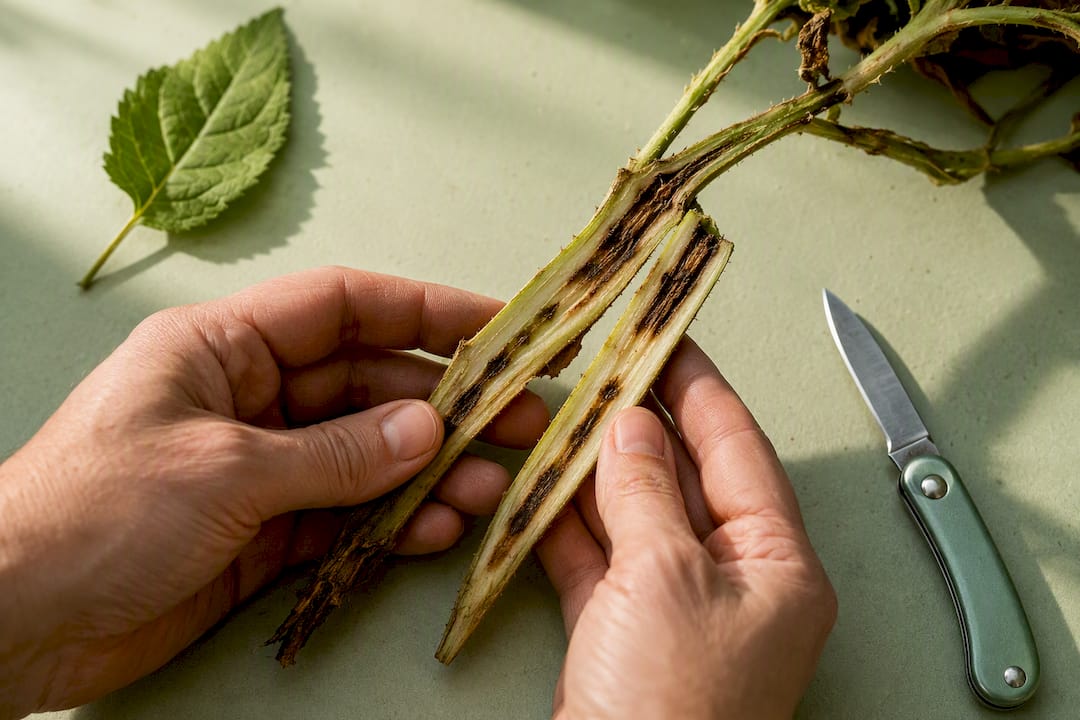
Pro Tip: Cut a symptomatic stem lengthwise and check the vascular tissue inside. Brown or black streaking in the water-conducting tissue is a classic sign of vascular wilt diseases like Fusarium or Verticillium, and it’s invisible from the outside.
Misdiagnosis usually happens at this exact step: someone sees yellowing leaves, assumes “disease,” and skips the search for actual evidence. Every diagnostic clinic that reviews home gardener submissions sees the same pattern repeated: a photo of a sick-looking plant with no close-up of the tissue that would actually confirm what’s wrong. Take the wide shot for context, then get in close. A macro lens attachment for a phone camera costs less than a bag of fertilizer and pays for itself the first time it saves you from spraying a fungicide on a problem that was actually bacterial (fungicides don’t touch bacteria) or abiotic (fungicides don’t touch anything, in that case).
If you genuinely can’t find a sign after a careful look, that’s information too. It shifts the probability toward an abiotic cause, an early-stage infection that hasn’t sporulated yet, or a pathogen that needs lab equipment to detect at all, which is a thread this article picks back up when it gets to testing.
How Do Distribution Patterns Reveal the Cause?
Where and when a problem shows up often narrows the list of suspects faster than the damage itself. A trained eye reads a garden bed the way a detective reads a room.
Start with distribution. If every plant in a row shows identical symptoms at the same growth stage, think environmental or cultural: irrigation line failure, a herbicide drift event, a soil pH problem, or a fertilizer burn from an uneven application. If damage clusters around field edges or fence lines, suspect drift from a neighboring application, salt spray from a road, or foot traffic compaction. Random, scattered plants with no obvious geographic logic point more toward an insect-vectored pathogen or a soil-borne organism with patchy distribution, since insects don’t feed in straight lines and root pathogens often exist in pockets tied to old planting holes or poor drainage.
Timing matters just as much as location. A few questions to run through as you observe:
- Did the problem appear right after a weather event? Heavy rain, hail, frost, or a heat spike can trigger symptoms that look infectious but are purely physiological.
- Did it follow a specific garden task? New mulch, a fresh application of compost, transplanting, or pruning can all introduce pathogens or trigger stress responses within days.
- Is it worse on one side of the plant? Sun-facing scorch, wind-facing desiccation, or damage concentrated on the side nearest a downspout all suggest environmental exposure rather than disease.
- Are nearby but different plant species affected too? A pathogen with a narrow host range hitting only one species points toward a specific disease; damage crossing unrelated species points toward something environmental, like contaminated irrigation water or a chemical spill.
Host range is an underused diagnostic tool. Powdery mildew on your squash and roses at the same time, in the same week, is almost certainly two different powdery mildew species, since most powdery mildews are host-specific. That kind of cross-referencing takes thirty seconds and rules out an entire category of explanation.
Pro Tip: Keep a simple photo log, dated and tagged by plant location, from the first day you notice anything unusual. A disease that spreads outward from a single point over two weeks tells a completely different story than one that appeared everywhere overnight, and you’ll only catch that difference if the timestamps exist.
What Questions and Samples Help You Diagnose Plant Disease?
A short, specific set of questions turns a vague “my plant looks sick” into something a clinic, an extension office, or an AI tool can actually work with. This is the same interrogation a professional diagnostician runs through before touching a microscope.
Ask yourself, or the person you’re helping:
- When did you first notice the problem, and how long has it been progressing?
- Is it spreading to nearby plants, staying contained, or improving on its own?
- What treatments, fertilizers, or pesticides have already been applied, and when?
- Has anything changed recently: new mulch, new soil, a repotting, a new watering schedule, construction nearby?
- Is this a plant variety with known disease susceptibilities, and where did it come from (nursery stock has its own risk profile versus a seed you started yourself)?
That last question matters more than most gardeners realize. Field history and variety pedigree are genuinely underrated diagnostic clues. A tomato variety with no resistance genes bred in is a completely different diagnostic starting point than a variety marked VFN (resistant to Verticillium, Fusarium, and nematodes) showing the same symptoms.
Once you’ve got answers, collect a sample the right way. For foliar problems, pull leaves showing early-to-moderate symptoms, not the ones already dead and dried out. Dead tissue is colonized by dozens of secondary organisms that have nothing to do with the original problem, and it muddies any lab result. For stem or vascular issues, cut a section that includes both healthy and symptomatic tissue so the transition zone is visible. For root problems, dig, don’t pull, so the root system arrives intact with some surrounding soil.
Sample from the active margin of a lesion, the zone where the pathogen is still actively spreading, according to the diagnostic framework APSnet lays out for working diagnosticians. That’s where you’ll find the highest concentration of the actual causal agent, versus tissue that’s already dead and colonized by whatever showed up second.
Photograph before you cut anything loose. Take a wide shot showing the whole plant and its surroundings, a medium shot of the affected area, and a close-up with something for scale (a coin, a ruler, your thumbnail) sitting next to the lesion. Photograph multiple leaves or stems at different stages of symptom development if you can find them on the same plant. A remote diagnosis from good photos and a solid history is often enough for common, well-known problems. It’s not enough when the symptoms are ambiguous, when the crop is high-value, or when you need a legally defensible identification for something like a regulated pathogen.
When Should You Send a Sample to a Plant Disease Lab?
Lab testing exists for the moment visual inspection runs out of answers, and knowing which test to request saves both time and money. The guiding principle at diagnostic clinics is blunt: test, don’t guess.
Different tests exist because different pathogens hide differently. Fungi that produce visible fruiting structures can often be identified under a microscope or grown out in culture, isolating the organism on a nutrient medium to watch how it grows. Bacteria get streaked onto selective media that favor their growth over contaminants. Viruses can’t be cultured at all in the traditional sense, so labs turn to ELISA (enzyme-linked immunosorbent assay), which detects viral proteins using antibodies, or PCR (polymerase chain reaction), which amplifies and detects the pathogen’s genetic material directly. Soil-borne nematodes get extracted from a soil sample and counted or identified under magnification in a dedicated nematode assay.
A 2015 review of plant disease detection methods makes the point that some pathogens simply cannot be grown in culture at all, which is why molecular and serological methods exist as a parallel track rather than a backup plan. Obligate pathogens that only survive inside living host tissue need PCR or ELISA because there’s no petri dish that will grow them.
A rough matching guide for which test fits which suspicion:
| Suspected cause | Best-suited test | What it detects |
|---|---|---|
| Fungal leaf spot or blight | Culture and microscopy | Fungal structures, spore morphology |
| Bacterial wilt or leaf spot | Selective culture, sometimes PCR | Bacterial colonies, species-specific DNA |
| Viral mosaic or stunting | ELISA or PCR | Viral proteins or viral genetic material |
| Root rot with no visible fungus | Soil assay, culture | Soil-borne fungal or oomycete pathogens |
| Suspected nematode damage | Nematode assay | Live nematodes extracted from soil/roots |
Submission logistics vary by clinic, but most university plant disease clinics, including Cornell’s, ask for a completed submission form describing symptoms and history, a properly packaged fresh sample (not mailed in a sealed plastic bag where it will rot in transit), and a modest per-sample fee similar to that of a home soil test. Turnaround for culture-based diagnosis usually takes several days to two weeks, since fungi and bacteria need time to grow out. PCR and ELISA results often come back faster, sometimes within a few days, because they don’t depend on culturing a living organism first.
When a pathogen is genuinely hard to culture, the practical move is to submit two portions of the same sample: one preserved and shipped for molecular testing, and one kept fresh, cool, and moist exactly as the clinic instructs for any live-culture attempt they still want to try. That hedges your bet across both detection strategies instead of betting everything on one.
Reading the result back is its own skill. A positive PCR result confirms the pathogen’s genetic material is present, but doesn’t always confirm it’s the primary cause of the symptoms you photographed. Some pathogens are opportunistic and show up on already-weakened tissue. A good lab report will note the confidence level and any secondary organisms found, and it’s worth asking the clinic directly if the report doesn’t make that clear.
How Do You Finalize a Plant Disease Diagnosis?
A defensible diagnosis needs your field observations and any lab result to agree, or at least not contradict each other in ways you can’t explain. Diagnosis, as APSnet frames it, is a cycle of hypothesis testing, not a single lookup: you observe, propose a cause, test that proposal, and revise if the evidence doesn’t fit.
When field observation and lab results line up, you’re done, and you can move straight to management. When they conflict, that’s a signal to dig further rather than pick whichever answer is more convenient. A lab result showing a common secondary fungus while your field notes point clearly to bacterial ooze and water-soaked lesions means the fungus is probably an opportunist on already-damaged tissue, not the primary cause.
While you wait on any test, take these steps immediately:
- Isolate the affected plant or bed if the pattern suggests something contagious, especially for anything showing bacterial ooze or a rapidly expanding lesion border.
- Disinfect tools between cuts on different plants using a diluted bleach solution or rubbing alcohol, since pruners are one of the most common ways gardeners spread pathogens themselves.
- Remove and discard (don’t compost) severely infected tissue for anything you already strongly suspect is infectious, since home compost piles rarely reach temperatures high enough to kill plant pathogens reliably.
- Hold off on broad-spectrum treatments until you know whether you’re dealing with fungal, bacterial, viral, or abiotic causes, since a fungicide does nothing for bacteria and both do nothing for a nutrient deficiency.
- Adjust the obvious cultural factor if one stands out, like overhead watering that’s kept foliage wet for hours each evening, a known driver of many fungal and bacterial leaf diseases.
Management differs sharply by category once confirmed; for targeted treatment workflows that link diagnosis to efficient intervention, consider Bugs Patrol Canberra - Professional Pest Control Services. Fungal diseases often respond to targeted fungicides, improved airflow, and removing infected debris. Bacterial diseases have far fewer effective chemical options and lean heavily on sanitation, resistant varieties, and avoiding overhead irrigation. Viral diseases have no cure at all once a plant is infected; management means removing infected plants to stop spread and controlling the insect vectors, like aphids or whiteflies, that carry many plant viruses between hosts. Abiotic problems get fixed by correcting the underlying condition: adjusting pH, changing watering habits, or moving a plant out of drift range.
Escalate beyond your own judgment when the stakes rise. A high-value ornamental tree, a commercial planting, or symptoms matching a regulated pest or pathogen warrants a call to your local extension office, a certified arborist, or a commercial diagnostic lab rather than a DIY call. Some plant diseases are reportable by law, and a professional will know which ones apply in your situation.
What Are the Visual Clues for Common Plant Diseases?
A fast triage pass through the major disease categories catches most home garden problems within minutes, before any lab is involved. Think of this as pattern matching against the most frequent suspects.
Fungal diseases tend to announce themselves visually. Powdery mildew shows up as a white to gray powdery coating on leaf surfaces, most common on squash, cucumbers, roses, and lilacs, and it thrives in humid conditions with poor airflow. Rust diseases produce orange, yellow, or reddish-brown pustules, often on leaf undersides, and they’re common across cereal crops, roses, and many ornamentals. Late blight, notorious on tomatoes and potatoes, produces dark, water-soaked lesions with a fuzzy white fungal growth visible at the lesion edge in humid weather, and it can defoliate a planting within days once conditions favor it.
Bacterial diseases look and behave differently even when the surface symptom seems similar. Water-soaked lesions that look almost translucent, sometimes surrounded by a yellow halo, suggest bacteria before fungus. The bacterial ooze test described earlier, cutting a stem and watching for a milky streaming discharge in water, is one of the most reliable field checks available for separating bacterial wilt from its fungal look-alikes. Bacterial diseases also tend to spread faster in wet, warm weather and often show angular leaf spots that follow leaf veins, unlike the rounder spots typical of many fungal infections.
Viral indicators are the category most likely to require lab confirmation, because visual clues overlap heavily with nutrient problems. Mottled or mosaic patterns of light and dark green on leaves, stunted growth that doesn’t respond to feeding, and distorted or narrowed leaf shapes (sometimes called “shoestring” symptoms) all point toward viral infection. No sign is visible for most plant viruses under normal field conditions, which is exactly why ELISA and PCR testing exist as the primary confirmation route rather than an afterthought.
Abiotic look-alikes trip up more gardeners than any actual disease. Nutrient deficiencies often show as patterned yellowing: interveinal chlorosis (yellowing between green veins) usually means iron or magnesium deficiency, while uniform yellowing starting on older leaves often points to nitrogen. Herbicide drift shows up as leaf cupping, twisting, or a bleached, streaky pattern, frequently on the side of the plant facing the source. Water stress, whether too much or too little, tends to affect the whole plant fairly uniformly rather than in the patchy or vein-following patterns typical of disease. A quick test for several of these: check soil moisture with a finger two inches down, and check whether damage correlates with wind direction from a recent spray application nearby.
How Does Viridia Fit Into a Plant Disease Diagnosis Workflow?
Photo-based health diagnosis tools work best as the first filter in the workflow described above, not the last word on it. Upload a photo of the affected plant, and AI can flag likely categories of concern based on visible symptoms, then provide suggested treatments matched to the plant’s needs and environmental factors.
Where it genuinely earns its place in the process:
- Rapid triage: a first-pass read on whether the issue is likely fungal, pest, or cultural, in seconds instead of hours of searching.
- Pattern tracking: logging symptoms over time in a garden journal, providing date-stamped records useful for spotting spread and progression.
- Recovery tracking: after beginning a treatment, checking back on the same plant to assess if the intervention is effective or if escalation is needed.
Here’s the honest limit: AI photo analysis supports triage and record-keeping, but it does not replace laboratory confirmation for viral pathogens, regulated diseases, or any case where a wrong guess is expensive. If Viridia’s diagnosis and your own field signs disagree, or if a treatment isn’t working after a reasonable trial period, that’s your cue to move to the lab-testing steps above.
Pro Tip: Export your photo log and notes from your garden journal before mailing a sample to a lab. A clean, dated record of when symptoms started and how they progressed gives the diagnostician real context instead of a single snapshot in time.
A Diagnostician’s Rule of Thumb
Treat every plant problem as a hypothesis you haven’t disproven yet, not a fact you’ve established. That mental shift alone would fix most of the misdiagnoses I see repeated across gardening forums: someone spots yellow leaves, decides it’s a disease, and starts treating before they’ve ruled out the three or four cheaper, more common explanations sitting right in front of them.
The habit that actually moves the needle isn’t expertise, it’s discipline. Date-stamp your photos. Keep a one-line log entry every few days. Write down what changed in the garden right before symptoms appeared. When two explanations still seem equally likely after all that, test rather than guess, because a wrong guess costs more in wasted product and lost time than a lab fee ever will.
None of this requires a plant pathology degree. It requires slowing down for about five extra minutes before reaching for a spray bottle, and treating your first theory as a starting point to test rather than a conclusion to defend.
— Dan
Speed Up Diagnosis and Track Recovery With Viridia
Photo-based tools can help get you from “something’s wrong with this plant” to a working theory in the time it takes to snap a photo, and they provide ongoing assistance after that first read. Point your camera at the affected leaf or stem, and a health diagnosis feature can check it against the plant’s identity, environmental factors, and suggest treatment paths.

The real value shows up after that first diagnosis. Every follow-up photo can be logged in a garden journal with a timestamp, allowing pattern tracking to happen automatically instead of remaining scattered in a camera roll. If a lab sample ends up being the next step, that dated history is exactly what a diagnostic clinic wants to see alongside a fresh tissue sample. Viridia won’t run a PCR test for you. What it will do is help you catch a problem early, document it properly, and know when it’s serious enough to escalate. Browse the plant care catalog for species-specific fiches once you’ve got a working diagnosis, or start with a custom garden plan to get ahead of recurring issues before next season.
Where to Go Deeper on Plant Disease Diagnosis
A handful of sources go well beyond what any single article can cover, particularly once you’re ready to submit a physical sample or want to see more reference photos.
- Cornell’s Plant Disease Diagnostic Clinic covers exact sample submission requirements and the clinic’s “Test, Don’t Guess” philosophy in detail.
- APSnet’s Plant Disease Diagnosis educational center walks through the full hypothesis-testing framework professionals use, with extensive visual references.
- The 2015 peer-reviewed review of detection methods is worth reading for anyone who wants the technical background on PCR, ELISA, and culture-based methods.
- Ohioline’s diagnostic factsheet from Ohio State University breaks the process down specifically for home gardeners working without lab access.
Sources
- Plant Disease Diagnostic Clinic | Plant Clinic services
- Plant Disease Diagnosis — APSnet Educational Center
- Current and prospective methods for plant disease detection — PMC (2015)
- Diagnosing sick plants — Ohioline (Ohio State University)